Female Pelvic Ultrasound Scan
Overview
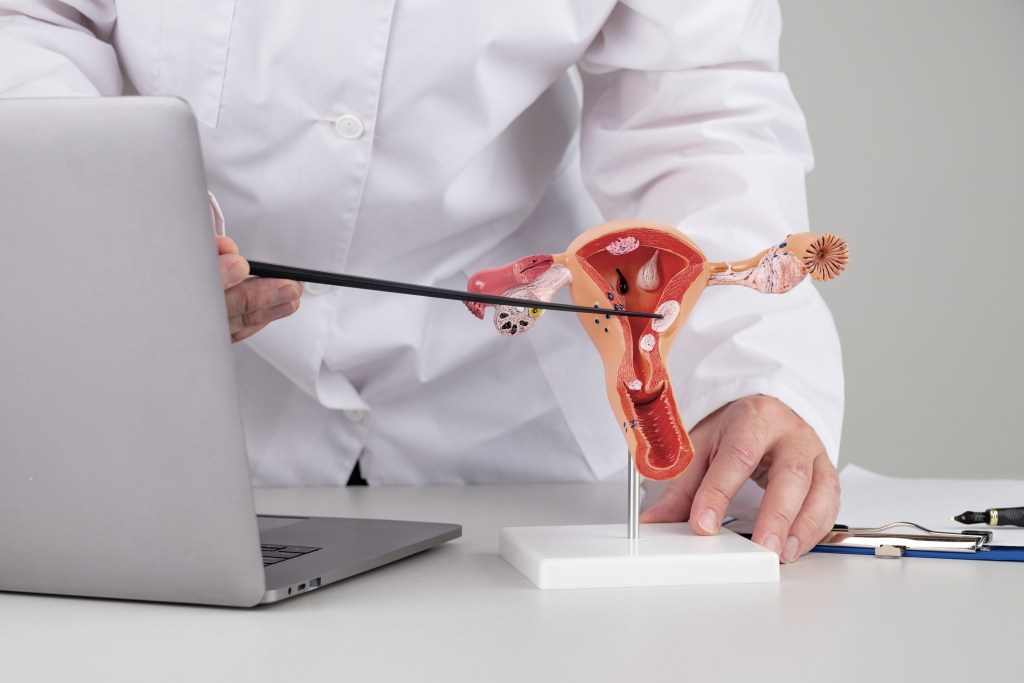
A pelvic ultrasound scan is a procedure that examines the health of the uterus and ovaries within the pelvic cavity. It is used to identify reasons for pain or discomfort and to detect early signs of potential issues.
Why have a pelvic ultrasound scan?
The most common reasons for pelvic ultrasound scan are pain, discomfort and abnormal bleeding. However, it may be possible to see conditions such as:
- Fibroids which are benign lumps of muscle in the uterus
- Adenomyosis (endometriosis in the uterus) which causes pain and abnormal bleeding
- Polyps which are found in the endometrium (cavity of the uterus) which are often the cause of abnormal bleeding
- Cysts on the ovaries which can cause a range of symptoms
- Endometriotic cysts on the ovaries.
It is rare for the above conditions to be harmful and most of the time no treatment is needed, some will even go away on their own. Sometimes it is a good idea to monitor these conditions as they can cause changes in the menstrual cycle. For example, fibroids can grow causing periods to become more painful and heavy, whilst polyps can cause intermittent bleeding. Cysts are a normal finding on the ovaries and they contain eggs. However, as women get older and they go through the menopause these cysts can become persistent and cause pain as well as irregularities in their periods.
- We can use ultrasound to look for abnormal shaped uteruses (congenital abnormalities). This is a condition that women are born with and are often incidental findings. However, they can affect fertility, cause heavy bleeding and it is important to know if this condition is present if you are considering pregnancy.
- We can check the position of your uterine coil whether it be a normal coil or a hormonal coil. This is important if you are experiencing pain or abnormal bleeding.
Ovarian Cancer
About 6,800 women are diagnosed with ovarian cancer in the UK each year and it has now become the fifth leading cause of death from cancer amongst women and its incidence is increasing. 5 out of every 100 cancers diagnosed in women are ovarian in nature. It is often termed “the silent killer” as it has no symptoms until it is well established in the body. Previously it was deemed to be a disease of post menopausal or older women, however, its incidence is increasing and it is becoming more common in pre-menopausal or younger women.
A transvaginal ultrasound scan can be used to look for signs of ovarian cancer. An early diagnosis allows for the correct treatment to be given thus preventing it from spreading to other organs in the body.
If during your scan at KMU we see something on the scan we are unhappy with we will ask your GP to do some blood tests and possibly refer you urgently to see a Gynaecologist for further tests.
Endometrial Cancer
Endometrial cancer is the fourth commonest cancer found in women. Unlike ovarian cancer, this cancer is often symptomatic, causing abnormal vaginal bleeding.
A transvaginal ultrasound scan can be used to help to see if the endometrium has an abnormal appearance and thickness. However, all post-menopausal women with abnormal vaginal bleeding should speak to their GP regardless of the scan results.
If during your scan at KMU we see something on the scan we are unhappy with we will ask your GP to do some blood tests and possibly refer you urgently to see a Gynaecologist for further tests.
How is a pelvic ultrasound scan carried out?
Transabdominal ultrasound – This is carried out by putting ultrasound gel on the skin surface of the abdomen. For this type of examination, you will need to have a full bladder which means drinking two pints of fluid one hour before you attend your examination. This is usually a less accurate way of examining the pelvic organs and is only suitable for those ladies who are unable to have a transvaginal scan.
Transvaginal ultrasound – This is an internal scan and for this examination you will not need to fill your bladder. This examination is usually painless and is routinely used at KMU, unless there is a contra-indication. It is the most detailed, sensitive and appropriate way to look at the uterus and ovaries. At KMU this examination is undertaken by female Sonographers who will be happy to discuss the examination with you if you have any concerns.
How do I get the results?
After the examination, the Sonographer will explain the findings and go through your report with you. You will receive two copies of the report, one for you and one for your doctor.
Limitations of ultrasound
Unfortunately, ultrasound has its limitations and we may not get the best views if you have a high BMI, excess gas in your pelvis, or if you haven’t followed the preparation instructions. If we think we can get better views in some of these cases we will rebook you for a follow-up scan, which will be free of charge.
Ultrasound can only detect abnormalities that are present at the time of the scan. Abnormalities can develop at any time, and while the Sonographer will look for early changes, they cannot predict the future. If you have concerns about anything, please discuss them with the Sonographer before, during or after the scan.
Annual pelvic assessment
KMU offers a yearly scan to check your uterus and ovaries at a reduced rate. This is so that we can monitor any changes in your pelvis and look for early signs of ovarian and endometrial cancer. We will notify you by post when your appointment is due if you wish to take up this offer.
